Detached Retina
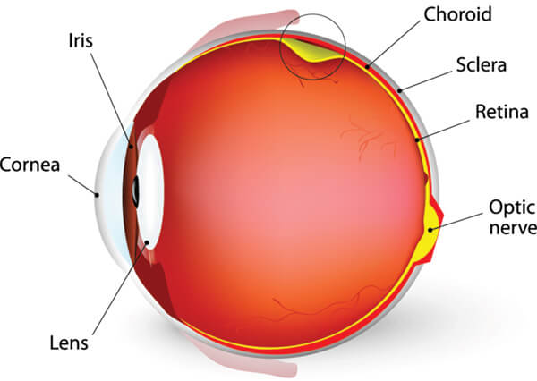
The retina is the light-sensitive tissue lining the back of our eye. Light rays are focused onto the retina through our cornea, pupil and lens. The retina converts the light rays into impulses that travel through the optic nerve to our brain, where they are interpreted as the images we see. A healthy, intact retina is key to clear vision.
The middle of the eye is filled with a clear gel called vitreous (vi-tree-us) that is attached to the retina. As we get older, the vitreous may shrink and pull on the retina. When this happens, you may notice what look like flashing lights, lightning streaks or the sensation of seeing “stars.” These are called flashes.
Usually, the vitreous moves away from the retina without causing problems. But sometimes the vitreous pulls hard enough to tear the retina in one or more places. Fluid may pass through a retinal tear, lifting the retina off the back of the eye — much as wallpaper can peel off a wall. When the retina is pulled away from the back of the eye like this, it is called a retinal detachment.
The retina does not work when it is detached and vision becomes blurry. A retinal detachment is a very serious problem that almost always causes blindness unless it is treated.
Symptoms
Symptoms of a retinal tear or a detached retina can include the following:
- A sudden increase in size and number of floaters, indicating a retinal tear may be occurring
- A sudden appearance of flashes, which could be the first stage of a retinal tear or detachment
- Having a shadow appear in the periphery (side) of your field of vision
- Seeing a gray curtain moving across your field of vision
- A sudden decrease in your vision
If you notice these symptoms, contact your OCB ophthalmologist immediately.
Risk Factors
People with the following conditions have an increased risk for retinal detachment:
- Nearsightedness
- Previous cataract surgery
- Glaucoma
- Severe eye injury
- Previous retinal detachment in the other eye
- Family history of retinal detachment
- Weak areas in the retina that can be seen by an ophthalmologist during an eye exam.
If you are very nearsighted or if you have a family history of retinal problems, be sure to have complete dilated eye exams on a regular basis. And always wear protective eyewear when playing sports or engaging in any other hazardous activities. If you have a serious eye injury, see your OCB ophthalmologist right away for an exam.
Treatment
Your OCB ophthalmologist can diagnose a retinal tear or retinal detachment during an eye examination where he or she dilates (widens) the pupils of your eyes. An ultrasound of the eye may also be performed to get additional detail of the retina.
Almost all patients with retinal detachments must have surgery to place the retina back in its proper position. Otherwise, the retina will lose the ability to function, possibly permanently, and blindness can result. The surgical method for fixing retinal detachment depends on the characteristics of the detachment. Your OCB ophthalmologist will discuss with you the type of procedure recommended for your retinal detachment that will give you the best possible outcome and will tell you about the various risks and benefits of your treatment options.